
 Radiology & Imaging has three radiologists specializing in cardiac CT and MRI. The division is directed by Christopher Moore, MD, Ph.D., and also includes Alena Kreychman, MD, and Yu Grace Zeng, MD. The program is strengthened by strong collaborations with Baystate Cardiology, as well as participation by cardiology fellows and upper-level radiology residents at the Baystate Medical Center.
Cardiac CT
Outpatient cardiac CT is performed at Baystate Medical Center, although coronary calcium scoring is also performed at the Baystate 3300 Main Street facility in Springfield, and the Baystate Radiology and Imaging outpatient facility at 325 King Street, Northampton.
The Most Common Reasons to Receive a Cardiac CT
- Coronary CT angiography
The most common reason for cardiac CT is to look for narrowing or blockage of coronary arteries. In some patients, this study may replace, or determine the need for, invasive catheter angiograms of the coronary arteries. This requires a relatively slow heart rate to avoid motion blurring, preferably 55-65 beats per minute, and often medicine is given by mouth and/or vein (such as metoprolol) to reduce heart rate. Imaging is timed to the heart cycle using an ECG. After a small injection of IV contrast to determine the timing of the scan, and usually, a nitroglycerine tablet under the tongue to enlarge the coronary arteries, a rapid injection of IV contrast is given and imaging of the heart is performed during a short breath-hold. Detailed images of the coronary arteries are then performed and evaluated. Some imaging protocols also allow wall motion to be assessed.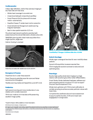
Download the PDF summary of the capabilities of the Revolution Apex CT Scanner.
- Coronary Calcium Scoring
The amount of calcification is measured in the coronary arteries to help determine a person’s risk for coronary artery disease or future heart attack. This exam is timed with the heart cycle using an ECG and is done in a short breath-hold. No contrast is needed.
- Pulmonary Vein Mapping
Some people with an abnormal heart rhythm called atrial fibrillation are candidates for a catheter-based treatment (ablation) to block abnormal conduction in the left atrium and adjacent pulmonary veins. This breath-hold exam uses IV contrast and provides the anatomy of the atrium and veins required for that procedure.
- Transcatheter Aortic Valve Replacement (TAVR)
Some patients with severe narrowing of the aortic valve (aortic stenosis), where the heart has difficulty pumping blood to the body, can have the valve replaced using catheters rather than open-heart surgery. The CT examination before the TAVR procedure is used to measure the size and shape of the valve region for proper selection of the replacement valve, and to assess the vessels that may be used for catheter access to the heart. This exam is timed with the heart cycle using an ECG and is done in a short breath-hold after IV contrast injection.
We Subspecialize In CT
At Radiology & Imaging, our radiologists subspecialize in one or more of the many types of CT scans. Your radiologist may primarily read and interpret chest CT scans or abdomen and pelvis CT scans. Or your radiologist may subspecialize in another part of the body–head, sinuses, or spine, for example. This means your images are analyzed by a physician with more experience, knowledge, and training.
Learn More
Visit our Links page for additional information.